
Last Updated: July 13, 2026
Introduction to the Familial Hypercholesterolemias
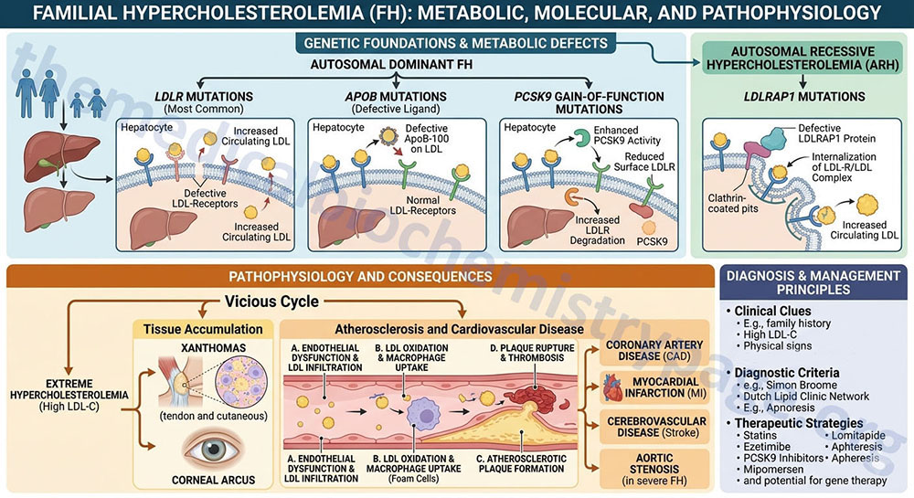
Classic familial hypercholesterolemia, FH (type 2a hyperlipidemia) is an autosomal dominant disorder that results from pathogenic variants affecting the structure and function of the cell-surface receptor that binds plasma LDL (low density lipoproteins) removing them from the circulation. The defects in LDL-receptor (LDLR) interaction result in lifelong elevation of LDL-cholesterol (LDL-C) in the blood. The resultant hypercholesterolemia leads to premature coronary artery disease and atherosclerotic plaque formation in the coronary arteries and the aorta. FH was the first inherited disorder that was recognized as being a cause of myocardial infarction (heart attack).
Although the primary causes of FH are pathogenic variants in the LDLR gene (representing 60%–80% of FH patients), pathogenic variants in the apolipoprotein B (APOB) gene that cause familial ligand-defective apoB (often referred to as familial hypercholesterolemia type 2) and gain-of-function variants in the proprotein convertase subtilisin/kexin type 9 (PCSK9) gene are also associated with autosomal dominant forms of familial hypercholesterolemia. Pathogenic variants in the APOB gene represent about 5%–10% of FH cases, whereas pathogenic variants in the PCSK9 gene represent less than 1% of identified cases of FH.
Another, very rare, form of familial hypercholesterolemia, that is inherited as an autosomal recessive disorder, results from pathogenic variants in the gene (LDLRAP1) that encodes the protein, low-density lipoprotein receptor adapter protein 1.
LDLR Pathogenic Variants and Familial Hypercholesterolemia
Although the disease is inherited in an autosomal dominant manner, the disease exhibits a gene dosage effect. Homozygous individuals are more severely affected than are heterozygotes. Heterozygotes for FH occur with a frequency of 1 in 500 which makes this disease one of the most common inherited disorders in metabolism.
The LDLR gene is located on chromosome 19p13.2 spanning 45 kilobases (kb) and is composed of 18 exons that generate five alternatively spliced mRNAs. These five mRNAs encode LDLR isoforms identified as isoform 1 (860 amino acids), 2 (858 amino acids), 3 (819 amino acids), 4 (692 amino acids), and 6 (682 amino acids).
Six different classes of receptor pathogenic variants have been identified in FH with each class having multiple alleles. More than 700 different pathogenic variants in the LDLR gene have been found in FH.
The class 1 pathogenic variants are null variants because these result in a failure to make any detectable LDLR protein.
Class 2 pathogenic variants are the most common and represent intracellular transport defects. The LDLR does not move between the endoplasmic reticulum (ER) and the Golgi membranes.
Class 3 pathogenic variants represent LDLRs that are delivered to the cell surface but fail in their ability to bind LDL.
Class 4 pathogenic variants are the rarest and result in LDLR protein that will bind LDL but the LDLR-LDL complexes cannot be internalized.
Class 5 pathogenic variants result in receptors that bind and internalize the LDL particle but cannot release the particles in the endosomes so the receptors cannot recycle back to the cell surface.
Class 6 pathogenic variants are in the cytoplasmic tail of the protein which leads to failure to reach the hepatocyte plasma membrane.
Clinical Findings in Familial Hypercholesterolemia
The clinical characteristics of FH include elevated concentrations of plasma LDL and deposition of LDL-cholesterol in the arteries, tendons and skin. Fat deposits in the arteries are called atheromas and in the skin and tendons they are called xanthomas. In heterozygotes, hypercholesterolemia is the earliest clinical manifestation in FH and remains the only clinical finding during the first decade of life. Xanthomas and the characteristic arcus cornea (whitish ring on the peripheral cornea) begin to appear in the second decade. The symptoms of coronary heart disease present in the fourth decade. At the time of death 80% of heterozygotes will have xanthomas.
The clinical picture is more uniform and severe in homozygotes. Homozygotes present with marked hypercholesterolemia at birth and it will persist throughout life. Xanthomas, arcus cornea, and atherosclerosis will develop during childhood in homozygotes. Death from myocardial infarction usually will occur before the age of 30 in homozygotes. Shown in the Table below are representative values of various plasma lipids in normal, heterozygote FH, and homozygote FH individuals. These values are not intended to be used as diagnostic and can vary greatly in certain individuals from those given here.
| Genotype | Age, yrs | Total Cholesterol (mg/dl) | LDL (mg/dl) | HDL (mg/dl) | Triglycerides (mg/dl) |
| Normal | 1-19 | 175 ± 30 | 110 ± 25 | 55 ± 15 | 60 ± 25 |
| Heterozygotes | 1-19 | 300 ± 60 | 240 ± 60 | 45 ± 10 | 80 ± 50 |
| Homozygotes | 1-19 | 680 ± 170 | 625 ± 160 | 35 ± 10 | 100 ± 50 |
| Normal | >20 | 200 ± 40 | 125 ± 30 | 55 ± 15 | 80 ± 30 |
| Heterozygotes | >20 | 380 ± 80 | 300 ± 80 | 45 ± 15 | 150 ± 75 |
LDLR Pathogenic Variants in Familial Hypercholesterolemia
As indicated above there have been over 700 different pathogenic variants in the LDLR gene identified in FH patients. Of diagnostic significance is the fact that 45 different polymorphisms in the LDLR gene have been identified and can be detected using RFLP and/or SSCP techniques.
Class 1 Pathogenic Variants
Multiple molecular mechanisms have been shown to result in the null variants that comprise the class 1 FH family. The alterations include deletions that eliminate the LDLR gene promoter. In addition, frameshift, nonsense and splicing variants cause the null phenotype.
Class 2 Pathogenic Variants
There are two subclasses of class 2 pathogenic variants in FH. Class 2A pathogenic variants result in an LDLR protein that fails to be transported out of the ER. Class 2B pathogenic variants are “leaky” in that some of the newly synthesized LDLR protein is transported to the Golgi but at a reduced rate compared to wild-type. The class 2B pathogenic variants are the more common type. Most of the class 2 pathogenic variants are clustered in the exons that comprise the LDL-binding domain. The LDLR protein has a domain with homology to the epidermal growth factor (EGF) precursor protein (see the Growth Factors page) and most of the rest of the class 2 pathogenic variants are found in this EGF precursor homology region.
Class 3 Pathogenic Variants
Most of the class 3 alleles result from in-frame rearrangements in the cysteine-rich repeats of the LDL-binding domain or in the EGF precursor domain. Because there is a similarity in the class 2 and class 3 pathogenic variants, it is difficult to assess which class of pathogenic variant is causing the observed FH phenotype. To accurately distinguish class 3 and class 2B alleles at the functional level it is necessary to isolate fibroblasts from the patient and do in vitro ligand-binding assays.
Class 4 Pathogenic Variants
All of the class 4 alleles have been shown to affect the cytoplasmic domain of the LDLR protein. In addition, class 4 alleles can be divided into two subclasses dependent upon whether the pathogenic variants affect only the cytoplasmic domain or include pathogenic variants in the adjacent membrane-spanning domain.
Class 5 Pathogenic Variants
Deletion or alteration of the EGF precursor homology domain results in class 5 alleles. These variant LDLR proteins do not recycle back to the plasma membrane following LDL-stimulated endocytosis. The total percentage of FH alleles that are of the class 5 type may be underestimated because the class 5 pathogenic variants can produce a phenotype that somewhat resembles that of class 3 pathogenic variants (i.e. deficient LDL binding).
Class 6 Pathogenic Variants
The class 6 pathogenic variants are associated with alterations in the cytoplasmic tail of the protein. This class of variant LDLR do not reach the liver cell membrane and are most likely rapidly degraded.
APOB Pathogenic Variants and Familial Hypercholesterolemia
The APOB gene is located on chromosome 2p24.1 and is composed of 29 exons that encode a 4563 amino acid precursor protein which is processed in the liver to apo-B100 (referring to 100% of the coding capacity). The mRNA encoded by the APOB gene is edited in the small intestines at nucleotide 2180 such that a CAA codon is converted to a UAA stop codon resulting in a shorter, 2179 amino acid precursor protein. This shorter intestinal-specific protein is designated apo-B48 since it is composed of 48% of the full-length (apo-B100) APOB mRNA encoded protein.
Pathogenic variants in the APOB gene are associated with two distinctly different pathologies. Pathogenic variants that affect the LDL receptor binding domain of apolipoprotein B-100 (apoB-100) result in a form of familial hypercholesterolemia referred to as familial ligand-defective apoB.
Pathogenic variants in the APOB gene that generate a truncation of the encoded protein result in a disorder termed familial hypobetalipoproteinemia (FHBL). APOB gene variants that lead to truncation of apolipoprotein B include nonsense, frameshift, and splice site errors. The characteristic features of FHBL are low serum levels (25%–35% of normal) of apolipoprotein B, LDL cholesterol, and total cholesterol.
The pathology associated with APOB gene variants that affect the LDL receptor-binding domain is similar to the pathology of LDLR pathogenic variants, namely severe elevations in serum LDL cholesterol and early onset coronary atherosclerosis. However, familial ligand-defective apoB patients usually have less severe hypercholesterolemia, reduced incidence of xanthomas, and slightly lower incidence of coronary artery disease.
Familial ligand-defective apoB results from four different pathogenic variants in the APOB gene. The most common pathogenic variant in the APOB gene found in this disorder results in the substitution of a glutamine (Gln; Q) for arginine (Arg; R) at amino acid 3500, identified as the R3500Q variant. Another pathogenic variant at the same codon results in substitution of the Arg for tryptophan (Trp; W), identified as the R3500W variant. The other two identified pathogenic variants alter codon 3531 resulting in substitution of cysteine (Cys; C) for Arg, identified as the C3531R variant, and codon 3480 where Trp is substituted for Arg, identified as the R3480W variant.
PCSK9 Pathogenic Variants and Familial Hypercholesterolemia
Rare causes of autosomal dominant familial hypercholesterolemia have been associated with gain-of-function variants in the gene (PCSK9) encoding proprotein convertase subtilisin/kexin type 9.
The PCSK9 gene is located on chromosome 1p32.3 and is composed of 14 exons that generate nine alternatively spliced mRNAs, each of which encode distinct preproprotein isoforms.
The normal function of PCSK9 is to degrade some of the LDL receptors that are endocytosed following binding of LDL. PCSK9 is a secreted protein that associates with the extracellular domain of the LDL receptor, predominantly on hepatocytes, and is co-internalized with LDL-LDL receptor complexes. The action of PCSK9 on LDL receptor protein in the endosomal compartment following internalization results in less than 100% of the LDL receptors being recycled to the plasma membrane. Therefore, PCSK9 pathogenic variants that enhance the activity of the enzyme will result in even greater level of LDL receptor degradation and dramatically less LDL receptor recycling. This will result in significant reduction in the capacity for LDL binding with a consequent elevation in serum LDL levels, typical of familial hypercholesterolemia.
Loss-of-function pathogenic variants in the PCSK9 gene result in hypocholesterolemia and a reduced risk for coronary artery disease. The hypocholesterolemia associated with loss-of-function pathogenic variants in the PCSK9 gene is similar to that observed in patients with familial hypobetalipoproteinemia (FHBL) that is classically the result of pathogenic variants in the APOB gene that result in the synthesis of truncated apolipoprotein B protein.
LDLRAP1 Pathogenic Variants and Familial Hypercholesterolemia
As indicated in the Introduction, a very rare form of familial hypercholesterolemia, that is inherited as an autosomal recessive disorder, results from pathogenic variants in the gene (LDLRAP1) that encodes the protein, low-density lipoprotein receptor adapter protein 1.
The LDLRAP1 gene is located on chromosome 1p36.11 and is composed of 15 exons that encode a 308 amino acid protein.
Low-density lipoprotein receptor adapter protein 1 is localized to the cytosol. The normal function of the protein is to bind to the tyrosine phosphorylated LDL receptor and facilitate endocytosis following the binding of LDL particles to the extracellular ligand binding domain.
The disorder that results from pathogenic variants in the LDLRAP1 gene is also referred to as autosomal recessive familial hypercholesterolemia, ARH. Similar to the other more common autosomal dominant forms of familial hypercholesterolemia, ARH is associated with a markedly elevated serum concentration of low-density lipoprotein (LDL), xanthomas, and premature coronary artery disease.