Last Updated: October 24, 2025
Introduction to Carbohydrates
Carbohydrates are carbon compounds that contain large quantities of hydroxyl groups. The simplest carbohydrates also contain either an aldehyde moiety (these are termed polyhydroxyaldehydes) or a ketone moiety (polyhydroxyketones). All carbohydrates can be classified as either monosaccharides, oligosaccharides or polysaccharides. Anywhere from two to ten monosaccharide units, linked by glycosidic bonds, make up an oligosaccharide. Polysaccharides are much larger, containing hundreds of monosaccharide units. The presence of the hydroxyl groups allows carbohydrates to interact with the aqueous environment and to participate in hydrogen bonding, both within and between chains. Derivatives of the carbohydrates can contain nitrogens, phosphates and sulfur compounds. Carbohydrates also can combine with lipid to form glycolipids or with protein to form glycoproteins.
Carbohydrate Nomenclature
The predominant carbohydrates encountered in the body are structurally related to the aldotriose glyceraldehyde and to the ketotriose dihydroxyacetone. All carbohydrates contain at least one asymmetrical (chiral) carbon and are, therefore, optically active. In addition, carbohydrates can exist in either of two conformations, as determined by the orientation of the hydroxyl group about the asymmetric carbon farthest from the carbonyl. With a few exceptions, those carbohydrates that are of physiological significance exist in the D-conformation. The mirror-image conformations, called enantiomers, are in the L-conformation.

Monosaccharides
The monosaccharides commonly found in humans are classified according to the number of carbons they contain in their backbone structures. The major monosaccharides contain four to six carbon atoms.
Table of Carbohydrate Classifications
| # Carbons | Category Name | Relevant examples |
| 3 | Triose | Glyceraldehyde, Dihydroxyacetone |
| 4 | Tetrose | Erythrose |
| 5 | Pentose | Ribose, Ribulose, Xylulose |
| 6 | Hexose | Glucose, Galactose, Mannose, Fructose |
| 7 | Heptose | Sedoheptulose |
| 9 | Nonose | Neuraminic acid, also called sialic acid |
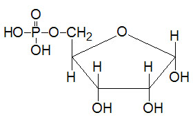
The aldehyde and ketone moieties of the carbohydrates with five and six carbons will spontaneously react with alcohol groups present in neighboring carbons to produce intramolecular hemiacetals or hemiketals, respectively. This results in the formation of five- or six-membered rings. Because the five-membered ring structure resembles the organic molecule furan, derivatives with this structure are termed furanoses. Those with six-membered rings resemble the organic molecule pyran and are termed pyranoses
Such structures can be depicted by either Fischer or Haworth style diagrams. The numbering of the carbons in carbohydrates proceeds from the carbonyl carbon, for aldoses, or the carbon nearest the carbonyl, for ketoses.
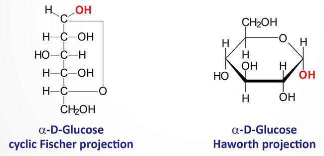
The rings can open and re-close, allowing rotation to occur about the carbon bearing the reactive carbonyl yielding two distinct configurations (α and β) of the hemiacetals and hemiketals. The carbon about which this rotation occurs is the anomeric carbon and the two forms are termed anomers. Carbohydrates can change spontaneously between the α and β configurations: a process known as mutarotation. When drawn in the Fischer projection, the α configuration places the hydroxyl attached to the anomeric carbon to the right, towards the ring. When drawn in the Haworth projection, the α configuration places the hydroxyl downward.
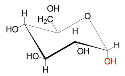
The spatial relationships of the atoms of the furanose and pyranose ring structures are more correctly described by the two conformations identified as the chair form and the boat form. The chair form is the more stable of the two. Constituents of the ring that project above or below the plane of the ring are axial and those that project parallel to the plane are equatorial. In the chair conformation, the orientation of the hydroxyl group about the anomeric carbon of α-D-glucose is axial and equatorial in β-D-glucose.
Disaccharides
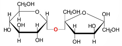
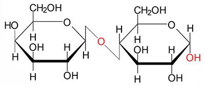
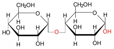
Covalent bonds between the anomeric hydroxyl of a cyclic sugar and the hydroxyl of a second sugar (or another alcohol containing compound) are termed glycosidic bonds, and the resultant molecules are glycosides. The linkage of two monosaccharides to form disaccharides involves a glycosidic bond. Several physiogically important disaccharides are sucrose, lactose and maltose.
Polysaccharides
Most of the carbohydrates found in nature occur in the form of high molecular weight polymers called polysaccharides. The monomeric building blocks used to generate polysaccharides can be varied; in all cases, however, the predominant monosaccharide found in polysaccharides is D-glucose. When polysaccharides are composed of a single monosaccharide building block, they are termed homopolysaccharides. Polysaccharides composed of more than one type of monosaccharide are termed heteropolysaccharides.
Glycogen
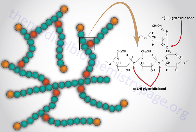
Glycogen is the major form of stored carbohydrate in animals. This crucial molecule is a homopolymer of glucose in α–(1,4) linkage; it is also highly branched, with α–(1,6) branch linkages occurring every 8-10 residues. Glycogen is a very compact structure that results from the coiling of the polymer chains. This compactness allows large amounts of carbon energy to be stored in a small volume, with little effect on cellular osmolarity.
Starch
Starch is the major form of stored carbohydrate in plant cells. Its structure is identical to glycogen, except for a much lower degree of branching (about every 20–30 residues). Unbranched starch is called amylose; branched starch is called amylopectin.

