Last Updated: September 15, 2025
Introduction to Marfan Syndrome
Marfan syndrome (MFS) is an autosomal dominant disorder affecting the connective tissue of numerous different organ systems. The characteristics of Marfan syndrome include flexible joints, long arms, legs, fingers, and toes. Most individuals with Marfan syndrome are tall and thin. The symptoms of MFS are the result of inherited defects in the gene encoding the extracellular matrix glycoprotein, fibrillin 1.
The manifestation of the various symptoms in multiple organ systems in individuals with Marfan syndrome defines the genetic phenomenon of pleiotropy. Pleiotropy is defined by the fact that a single genetic variant can influence multiple phenotypic traits.
Molecular Biology of Marfan Syndrome
Humans express three genes that encode members of the fibrillin family, FBN1, FBN2, and FBN3 which encode the proteins identified as fibrillin 1, fibrillin 2, and fibrillin 3, respectively.
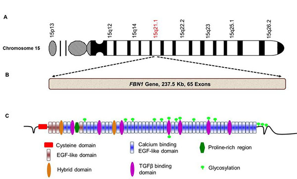
The FBN1 gene is located on chromosome 15q21.1 and spans around 200kb. The FBN1 gene is composed of 66 exons that generate four alternatively spliced mRNAs that collectively encode three distinct protein isoforms. The primary FBN1 encoded protein is a preprofibrillin of 2,871 amino acids with a processed molecular weight of approximately 350,000 Da.
There are five distinct structural regions in the fibrillin protein. The largest of these structural regions comprises 75% of the total protein and is composed of 46 EGF-like repeats. These EGF-like repeats are cysteine-rich domains that were first identified in the epidermal growth factor (EGF) protein. Almost all of the EGF-like domains are encoded by individual exons. There are eight additional cysteine motifs that share homology to a domain first identified in the latent transforming growth factor-β1-binding protein (LTBP1) called a TB domain. The fibrillin 1 protein has also been shown to bind calcium ions.
In addition to the defects in connective tissue, there are systemic effects in the cardiovascular, musculoskeletal, and ophthalmic systems as well as in the integument in Marfan patients.
As described below there are certain diagnostic criteria that allow for differentiating MFS from several related disorders such as congenital contractural arachnodactyly which is caused by mutations in FBN2 gene.
Functions of the Fibrillin 1 Protein
Fibrillin monomers link head to tail, as well as laterally, to generate microfibrils which can then form two and three dimensional structures. The most abundant fibrillin in elastic fibers is the FBN1 encoded protein, fibrillin 1. Fibrillin 1 serves as the scaffold in elastic fibers upon which cross-linked elastin is deposited. Although the functions of fibrillin 1 and fibrillin 2 are fairly well characterized the functions of fibrillin 3 are less well known. This discussion will focus on the functions known for fibrillin 1.
The fibrillins are large glycoproteins that complex with proteins of the latent TGFβ binding protein (LTBP) family and the fibulins into a structurally related family of extracellular matrix (ECM) proteins. There are four members of the LTBP family, LTBP-1, -2, -3, and -4.
The fibrillin-rich microfibrils play an important role in the extracellular regulation of endogenous TGFβ activity by providing a structural platform that controls the diffusion, storage, presentation, and release of several TGFβ family member proteins, particularly TGFβ-1, -2, and -3. Several other extracellular proteins that interact with fibrillin microfibrils, including small leucine-rich proteoglycans (LRPG), fibulin-4, microfibril-associated glycoprotein 1 (MAGP-1), emilin-1, and lysyl oxidase, are implicated in modulating TGFβ bioavailability and signaling.
Clinical Features of Marfan Syndrome
The cardinal manifestations of MFS are tall stature with dolichostenomelia (condition of unusually long and thin extremities) and arachnodactyly (abnormally long and slender fingers and toes), joint hypermobility and contracture, deformity of the spine and anterior chest, mitral valve prolapse, dilatation and dissection of the ascending aorta, pneumothorax and ectopia lentis (displacement of the crystalline lens of the eye). Correct diagnosis of MFS relies on the presence of a combination of manifestations in several organ systems as described below.
Skeletal Abnormalities
Individuals with MFS are taller at all ages than would be predicted based on their kinship. The tall stature is the result of overgrowth of the long bones which leads to the characteristic features of MFS such as disproportionately long legs, arms and digits. As indicated above this phenotype is called dolichostenomelia where one diagnostic feature is that the arm span will exceed the body height. The overgrowth of the tubular bones leads to deformity of the anterior chest. The can result in the ribs pushing the ribs either in (pectus excavatum) or out (pectus carinatum) or in on one side and out on the other. The skull is often elongated (termed dolichocephaly), the mandible underdeveloped with the palate narrow and highly arched, and the face is long and narrow. Because of ligament laxity the joints are hypermobile and there is often joint pain. Persons with MFS are now living longer due to clinical interventions in the cardiovascular abnormalities but this leads to an increased occurrence of degenerative skeletal problems such as osteoarthritis.
Cardiovascular Abnormalities
Mitral valve prolapse (MVP) is the most common finding in children with MFS. The prolapse is due to redundancy of the valve tissue, elongation of the valve annulus (valve ring) and elongation of the chordae tendineae (these are the tendons that connect the papillary muscles to the tricuspid and mitral valves). MVP will progress to severe mitral regurgitation which requires surgical intervention usually before 10 years of age. Progressive dilation of the proximal portions of both the aorta and main pulmonary arteries occurs in MFS and are highly important diagnostic and prognostic indicators.
Pulmonary Abnormalities
The combination of the deformity of the anterior chest and pectus excavatum results in a reduction in lung volume which can lead to severe restrictive pulmonary deficits. Spontaneous pneumothorax occurs in 4%–5% of MFS patients. Due to laxity of the hypopharyngeal tissue obstructive sleep apnea is a common symptom in MFS.
Ocular Abnormalities
The ocular hallmark of MFS is displacement of the lens from the center of the pupil (ectopia lentis). Because of the presence of this symptom in a high percentage of MFS patients, displacement of the lens in the absence of any traumatic event should lead to a consideration of MFS. The cornea of MFS patients is often flatter than normal. Detection of the corneal abnormality and surgical correction are necessary at an early age to prevent persistent amblyopia (lazy eye). Later in life many MFS patients will suffer from cataracts and/or glaucoma.

